
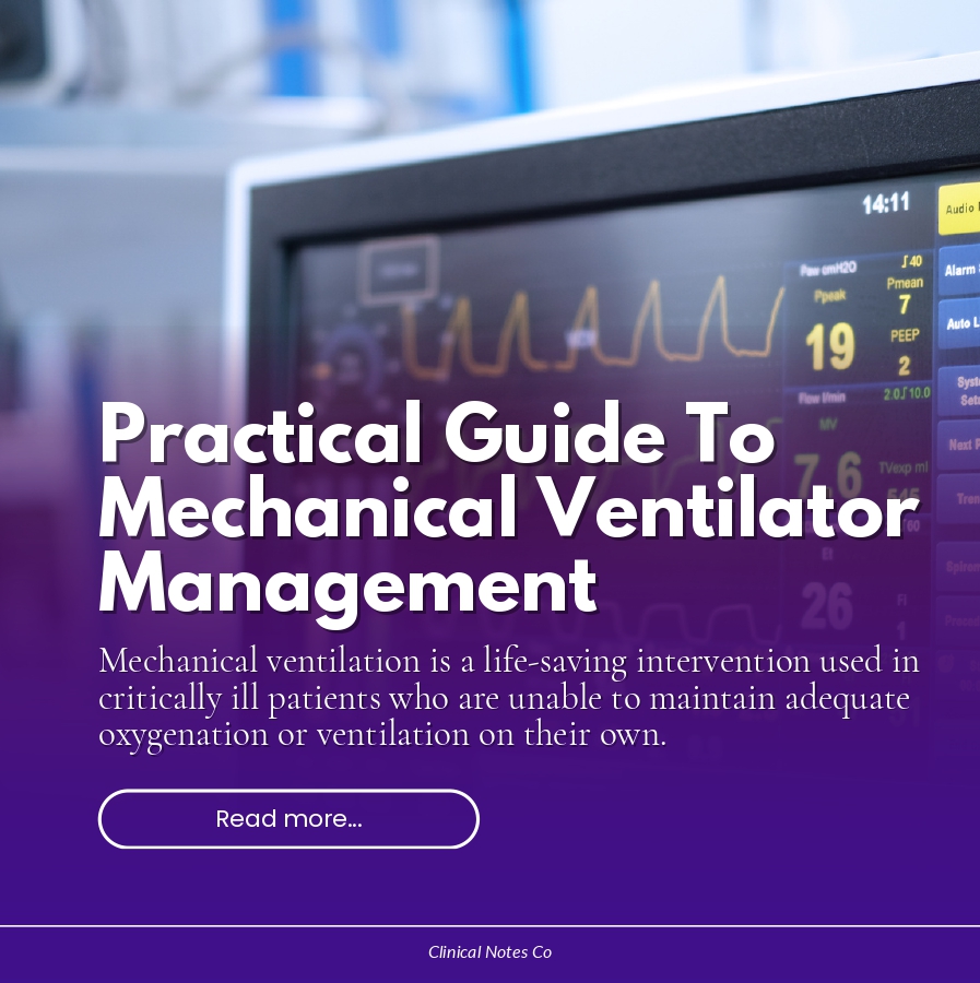
Mechanical ventilation is a life-saving intervention used in critically ill patients who are unable to maintain adequate oxygenation or ventilation on their own.
Effective ventilator management requires a solid understanding of respiratory physiology, ventilator modes, settings, and vigilant patient monitoring.
This blog provides a clear and practical overview of mechanical ventilator management, particularly useful for Respiratory therapist students, Nursing students, and healthcare professionals working in acute and critical care settings.
What is Mechanical Ventilation?
Mechanical ventilation is the use of a machine (ventilator) to assist or fully replace spontaneous breathing. It can be delivered invasively through an endotracheal tube or tracheostomy, or non-invasively via masks (e.g, BiPAP or CPAP)
Main goals of mechanical ventilation:
- Maintain adequate oxygenation
- Ensure effective ventilation (CO2 removal)
- Reduce work of breathing
- protect the lungs from further injury
Indications for Mechanical Ventilation
- Apnea
- Acute respiratory failure (hypoxemic or hypercapnic)
- Severe pneumonia or ARDS
- COPD exacerbation
- Neuromuscular disorders (e.g., Guillain-Barre syndrome)
- Decreased level of consciuosness
- Post-operative respiratory support
Ventilator Modes
1. Assist- Control (A/C)
- Each breath is either patient-triggered ot time-triggered
- Commonly used mode of ventilation
- The patient may initiate as many ventilator breaths as required above the set rate; therefore, the patient’s VE is not consistent
2. Synchronized Intermittent Mandatory Ventilation (SIMV)
- Allows for spontaneous breathing along with positive pressure ventilator breaths. it senses whent he patient is breathing spontaneously; therefore, no “breath stacking”occurs.
- It is used as both a weaning technique and for ventilation before weaning
3. Pressure Support Ventilation (PSV)
- This type of ventilation aids in the weaning process from the ventilator
- It is a patient-triggered, pressure-limited, flow-cycled breath, which maybe augmented with SIMV or used by itself in the CPAP mode
- It is used to make spontaneous breathing through the ET tube during weaning more comfortable by overcoming the high resistance and increasing inspiratory work caused by the ET tube ( 5 to 10 cm H2O is all that is required to overcome tubing resistance)
- An inspiratory pressure is set (usually 5 to 10 cm H2O for weaning purposes). As the patient initiates inspiration, the preset pressure is reached and held constant until a specific inspiratory flow is reached. then the pressure is terminated.
- The inspiratory pressure level may be set to achive a specific VT
4. Continuous Positive Airway Pressure (CPAP)
- May be achieved with the use of CPAP mask, nasal prongs, or intubation and a ventilator
- A preset pressure is maintained in the airways and alveoli as the patient breathes totally on his or her own. No positive pressure breaths are delivered.
- Patients whose PaO2 level cannot be maintained within normal limits using a 50% to 60% or more O2 mask and who have normal or low PaCO2 levels should be placed on CPAP. CPAP is also indicated for patients with obstructive sleep apnea who gainbenefit form the positive airway rpessure, which relieves the obstruction in the upper airway
- CPAP setups should always have a low-pressure alarm so that leaks in the system are detected
Ventilator Settings
1. Tidal Volume (VT)
- usually set at 6-8 mL/kg of ideal body weight
- Lower tidal volumes are used in ARDS to prevent lung injury
2. Respiratory Rate (RR)
- Determines minute ventilation
- adjusted based on PaCO2 levels
3. Fraction of Inspired Oxygen (FiO2)
- Percentage of oxygen delivered to the patient
- Aim to keep FiO2 lesser than or equal to 60% when possible to avoid oxygen toxicity
4. Positive End-Expiratory Pressure (PEEP)
- Used to maintain positive pressure in the airway after a ventilator breath
- Excessive PEEP levels may lead to decreases in PaO2 and lung compliance by over-distending already open alveoli and shunting blood to collapsed alveoli
OPTIMAL PEEP: The level of PEEP that improves lung compliance (CL) without cardiac compromise
a. Indications for PEEP
- Atelectasis
- PaO2 of lesser than 60 mmhg on FiO2 of greater than 50% O2
- Decreased functional residual capacity
- Decreased Lung compliance
- Pulmonary edema
b. Hazads of PEEP
- Barotrauma
- Decrease venous return
- Decreased cardiac output
- Decreased urinary output
5. Inspiratory flow control
- Normal setting: 40 to 80 L/min
- Adjusting the flow rate alters the inspiratory time, therefore altering the I:E ratio
6. I:E ratio
- A comparison of the inspiratory time with the expiratory time
- established by the use of three ventilator controls for volume controlles ventilation
Normal I:E ratio for the adults is 1:2
Normal I:E ratio fo infants 1:1
7. Sensitivity Control
- Determines the amount of patient effeort required to trigger the ventilator into inspiration
- Should be set to the patient generates 0.5 to 2.0 cmH2O pressure. This is referred to as pressure triggering
Comments ()