
Arterial Blood Gases (ABGs) can look intimidating at first—numbers, arrows, acid–base jargon—but once you understand the pattern, ABG interpretation becomes a powerful tool in patient assessment. Whether you’re working in ICU, ER, or med-surg, ABGs help you quickly evaluate oxygenation, ventilation, and acid–base balance.
Let’s break it down step by step.
What Are ABGs and Why Are They Important?
ABGs measure the effectiveness of the lungs and kidneys in maintaining homeostasis. They help answer key clinical questions such as:
- Is the patient ventilating adequately?
- Is oxygenation sufficient?
- Is there an acid-base imbalance?
- Is the condition respiratory or metabolic?
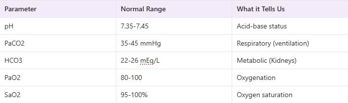
Components of ABGs (Normal Values)
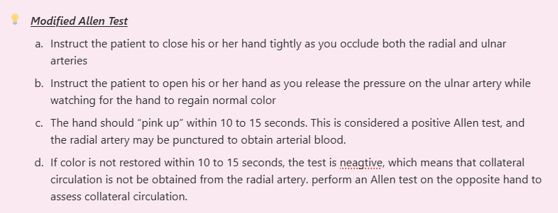
Sites from which to obtain arterial blood
1. Radial Artery
- Located in the wrist on the radial side (thumb side) close to the skin
- preferred site for sampling, not near to any large veins
- insertion of needle is at 45-degree angle
2.Brachial Artery
- Located on the medial side of the fossa near the insertion of the biceps
- Large and easily palpated
- No collateral circulation pressent, Close to a large vein and nerve
- Insertion of needle is at 60-degree angle
3. Dorsalis Pedis Artery
- Pertaneous cannulation is easy to perform and is relatively safe
- Incidence of thrombosis is less
- Effective collateral circulation exists with the posterior tibial artery
4. Femoral Artery
- Palpated laterally from the pubis bone
- A very large artery that can be easily palpated and presents a large target
- lacks collateral circulation
- Near a major vein (SCIATIC NERVE)
- Maybe the only site available in cases such as hypovolemia, hypotension or low cardiac output
- Insertion of needle is at a 90-degree angle
Arterial Oxygenation
1. PaO2
- The PaO2 is the portion of O2 that is dissolved in the plasma of the blood. For every 1 mmHg of PaO2 there is 0.003 mL of dissolved O2.
- The majority of O2 carried in the blood is bound to Hb
Normal Range: 80-100mmHg— ADULT
Normal PaO2 Value for NEONATES: 40-70 mmHg
Computation of PaO2 for ADULTS >60 years old: 80-(Age-60)= Min. baseline PaO2
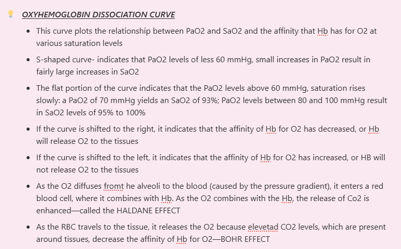
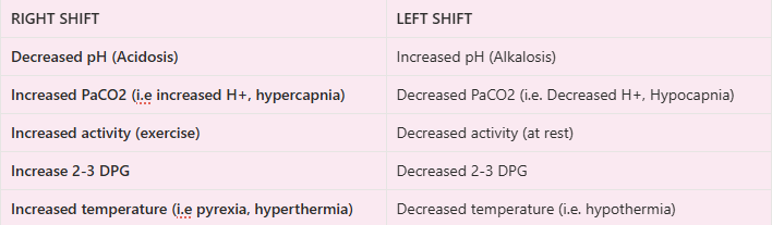
Factors Affecting Shifts of the HBO2 Curve
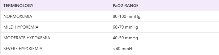
LEVELS OF HYPOXEMIA

2. SaO2 (Arterial Oxygen Saturation)
- The quality of O2 being carried by the Hb compared with the maximum that may be carried
Normal SaO2- greater than equal 97%
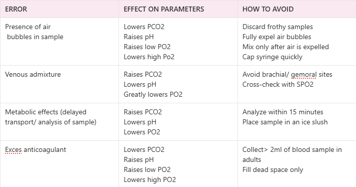
Preanalytical errors in ABG sampling
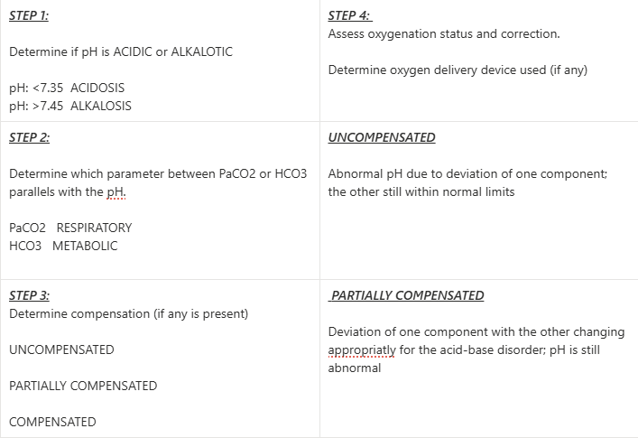
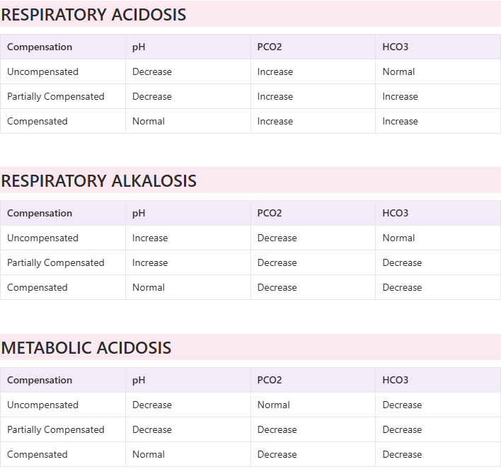
ARTERIAL BLOOD GAS INTERPRETATION
Patient at Room Air (21% Fio2)
Patient with Oxygen Therapy

RESPIRATORY DISTURBANCES
1. RESPIRATORY ACIDOSIS- primary causes by ALVEOLAR HYPOVENTILATION
- CNS depression (anesthesia, sedative drugs, narcotic analgesics)
- Airway obstruction (CO2 retention) (COPD, airway diseases)
- Neuromuscular disease (poliomyelitis, GBS, Myasthenia gravis)
- Trauma (severe restrictive disorders, kyphoscoliosis, pickwickian syndrome)
CORRECTION: Improve alveolar ventilation
2.RESPIRATORY ALKALOSIS- primary caused by HYPOXEMIA, which causes neural structures to increase ventilation
- Hyperventilation (anxiety, fear, stimulatory drugs, pain, CNS injuries)
- Stimulation of irritant receptors (pneumonia, pulmonary edema)
CORRECTION: Remove stimulus causing hyperventilation
3. METABOLIC ACIDOSIS
Can occur in two ways:
- Fixed acid accumulation in the blood
- Tissue hypoxia and anaerobic metabolism producing lactic acid
- Renal failure
- Ketoacidosis
- Ingestion of acids (oxalic acid, formic acid, aspirin intoxication)
b. Exessive loss of HCo3
- Severe diarrhea
- Ingestion of ammonium chloride
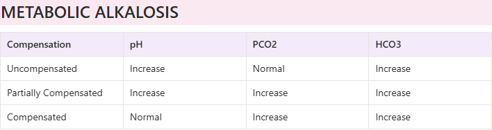
4. METABOLIC ALKALOSIS
Can occur in two ways:
- Loss of fixed (hydrogen ions)
- Vomiting
- Nasogastric drainage
- Diuretics
- Hypochloremia
- Hypovolemia
- Hypokalemia
b. Gain of blood buffer base
- Ingestion or infusion of HaHCO3
Comments ()