
Magnetic Resonance Imaging (MRI) plays a crucial role in the diagnosis, characterization, and monitoring of various pathologies. When it comes to chest thymoma, a pathology with significant clinical implications, the differentiation between T1-weighted (T1) and T2-weighted (T2) MRI sequences becomes highly relevant. In this article, we will explore the importance of T1 VS T2 MRI image in assessing chest thymoma and highlight the utility of each sequence.
- Anatomical Detailing with T1-Weighted Imaging
T1-weighted images predominantly provide detailed anatomical information about the scanned region. In the case of chest thymoma, T1 images can offer precise details about the tumor's size, margins, and its relationship to adjacent structures (1). This aids clinicians in understanding the tumor's exact position and its potential invasion into nearby tissues.
Reference:
Sakai S, Murayama S, Soeda H, et al. Thymic lesions: differentiation of thymic carcinoma from noninvasive thymoma, lymphoma, and mediastinal germ cell tumor at MR imaging. Radiology. 2001;221(1):191-196.
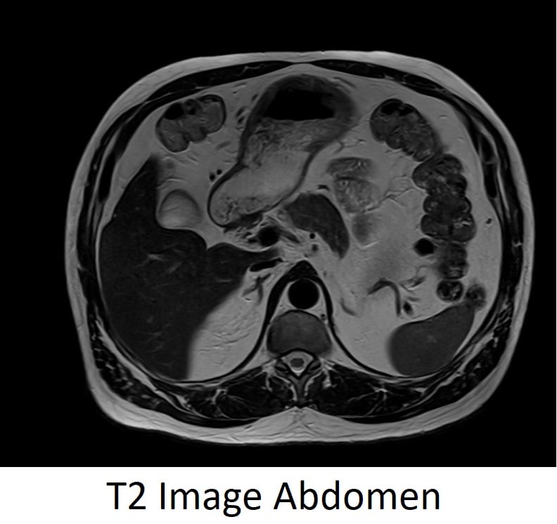
- Tumor Characterization with T2-Weighted Imaging
T2-weighted images are crucial for tissue characterization. Due to their heterogeneity, thymomas can display varying intensities in T2 images, enabling their differentiation from other mediastinal tumors or cysts. Additionally, T2 sequences can indicate the presence of necrotic or cystic components within the thymoma (2).
Reference: Tomiyama N, Müller NL, Ellis SJ, et al. Invasive and noninvasive thymoma: distinctive CT features. Journal of Computer Assisted Tomography. 2001;25(3):388-393.
- Detection of Fat Components
T1-weighted MRI sequences are sensitive to fat. Some thymomas, particularly those with lipid-rich content, appear hyperintense on T1-weighted images. Recognizing this pattern is important as it helps differentiate thymomas from other pathologies or provides insights into the tumor's histological subtype (3).
Reference: Inaoka T, Takahashi K, Mineta M, et al. Thymic hyperplasia and thymus gland tumors: differentiation with chemical shift MR imaging. Radiology. 2007;243(3):869-876.
- Evaluation of Tumor Invasion
Thymomas can potentially invade surrounding structures such as the chest wall, phrenic nerves, and great vessels. T2-weighted imaging, with its high contrast resolution, is instrumental in evaluating the extent of such invasive behavior, guiding subsequent therapeutic approaches (4).
Reference: Aquino SL, Kee ST, Warnick P, et al. Diagnosis of anterior mediastinal masses with computed tomography: pitfalls and pearls. Chest. 1999;116(1):17-22.
- Post-Treatment Monitoring
Following therapeutic interventions like surgery or chemotherapy, both T1 and T2 MRI sequences provide substantial data on tumor response, recurrence, or potential complications. Monitoring treatment outcomes is vital to ensure patient health and guide future therapeutic choices (5).
Reference: Marom EM, Milito MA, Moran CA, et al. Computed tomography findings of thymic carcinoma. Journal of Computer Assisted Tomography. 2001;25(6):905-910.
Conclusion
Both T1 and T2 MRI sequences hold significant diagnostic value in assessing chest thymoma. Their combined use offers a comprehensive view of the tumor, providing critical information for diagnosis, treatment planning, and post-treatment monitoring. As imaging technology continues to evolve, the precision and utility of MRI in thymoma assessment are expected to further enhance, underscoring the importance of understanding and leveraging both T1 and T2 sequences.