
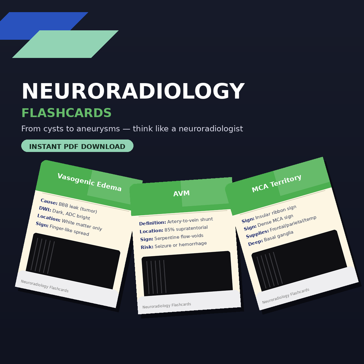
Neuroradiology Flashcards
On Sale
$8.99
$8.99
🧠 Neuroradiology Flashcards — Master Brain Imaging From Cysts to Aneurysms
The ultimate visual companion for neuroradiology — built for radiology residents, neurology trainees, and anyone who needs to recognize brain pathology fast and confidently.
Neuroradiology is one of the hardest subspecialties to master because so much rides on pattern recognition — a subtle signal difference, a location, a shape.
This flashcard deck breaks down the classic comparisons and high-yield entities that separate confident readers from guessers, paired with real annotated MRI and CT images.
🧠 What's Inside
Each card gives you:
- Overview & demographics — who gets it, and why it matters
- Key imaging characteristics — the sequence-specific findings that clinch the diagnosis
- Head-to-head comparisons — the differentiators examiners love to test
- Real annotated imaging — MRI (T1, T2, FLAIR, DWI/ADC, SWI) and CT examples
🩻 Topics Covered
Differentials & Pattern Recognition
- Epidermoid Cyst vs. Arachnoid Cyst
- Cytotoxic vs. Vasogenic Edema
- CNS Lymphoma vs. Toxoplasmosis
- Extra-axial vs. Intra-axial Mass Characteristics
- DWI & ADC Patterns (Restricted, Free Diffusion, T2 Shine-Through, T2 Black-Through)
Herniation Syndromes
- Subfalcine, Transtentorial (Uncal), and Cerebellar Tonsillar Herniation
Brain Tumors — Adult & Pediatric
- Low-grade → Glioblastoma (WHO Grade I–IV Astrocytoma spectrum)
- Juvenile Pilocytic Astrocytoma, Oligodendroglioma, Ganglioglioma, PXA
- Gliomatosis Cerebri, Choroid Plexus Papilloma/Carcinoma
- Intraventricular Meningioma, SEGA, Subependymoma
- Atypical Teratoid/Rhabdoid Tumor (AT/RT), Hemangioblastoma
- Common Intra-axial Tumors in Adults vs. Pediatrics (with mnemonics)
- Posterior Fossa Masses by age group
- Brain Lesion Complications — the "3 H's" (Hemorrhage, Hydrocephalus, Herniation)
Vascular Territories & Stroke
- ACA, MCA, PCA, and Anterior Choroidal Artery territories
- Watershed (Borderzone) Infarcts
- Vertebrobasilar System (PICA, AICA, SCA, Basilar perforators)
Vascular Malformations
- Arteriovenous Malformation (AVM)
- Cavernous Malformation (Cavernoma)
- Developmental Venous Anomaly (DVA)
- Capillary Telangiectasia
Aneurysms
- Saccular (Berry), Fusiform, Mycotic (Infectious) — types, causes, and imaging clues
Other Findings
- Normal Pressure Hydrocephalus (NPH) — triad, callosal angle, DESH sign
- Blooming Artifacts on SWI (hemorrhage, microbleeds, calcification)
- CNS Lymphoma imaging patterns (eccentric target sign, perfusion characteristics)
🎯 Who This Is For
- Radiology residents preparing for board exams
- Neurology and neurosurgery trainees
- Medical students on neuro or radiology rotations
- Anyone building pattern-recognition fluency for brain imaging
Format: PDF
Pages: 46 pages