

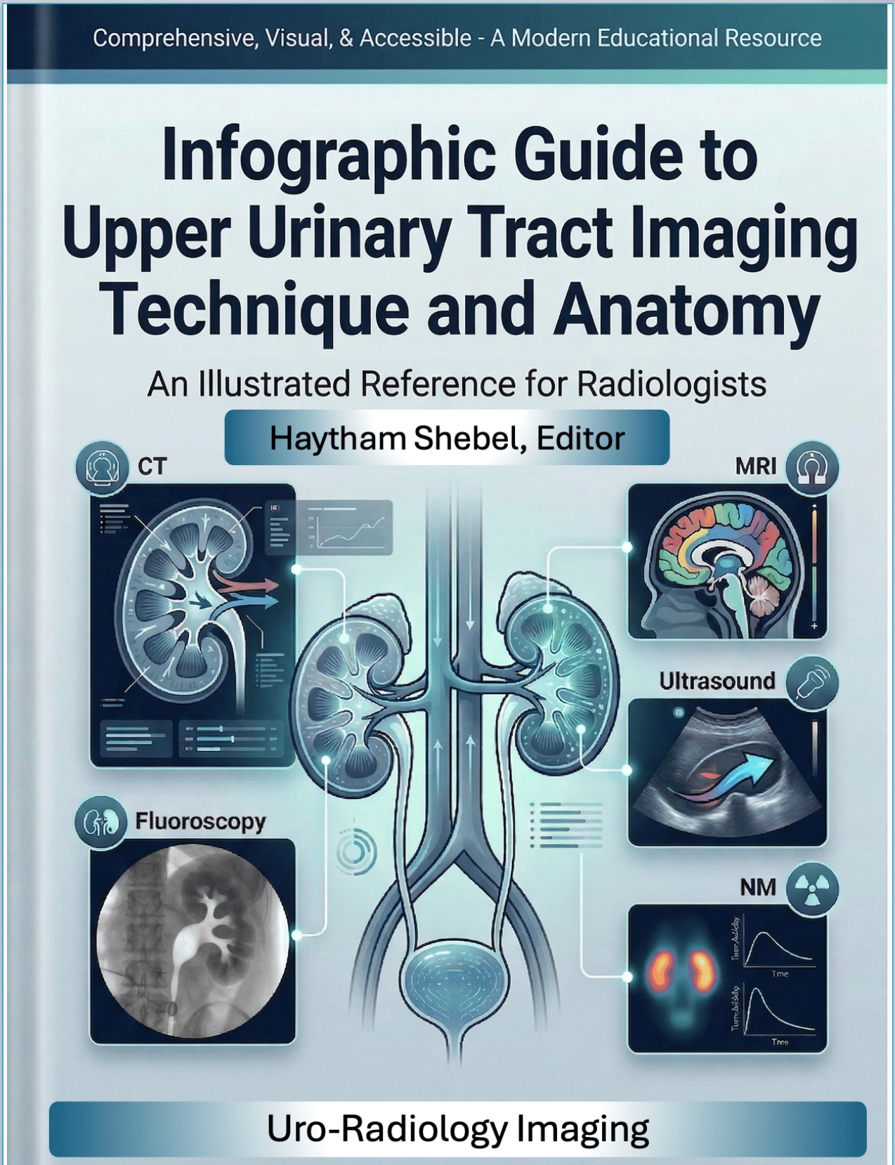
Infographic Guide of the Upper Urinary Tract Imaging
Infographic Guide to Upper Urinary Tract Imaging Technique and Anatomy By Prof. Haytham Shebel, MD Resource Author [Urogenital Section] of the Royal College of Radiologists [RCR],European Diploma in Urogenital Radiology (EDIUR) | ESUR Active Member | Certified Biomedical Research Harvard Medical School.
Upper urinary tract imaging is a technically demanding and anatomically rich subspecialty yet most available references present it through dense text, fragmented diagrams, and protocol tables that are difficult to retain and even harder to apply at the workstation.
This guide takes a fundamentally different approach.
Every concept from anatomical demonstration that include [whole Kidney, Kidney segmentation, Renal parenchyma, collecting system, Ureter, and retroperitoneum] and different imaging modalities from simple x-ray and IVU tecnhniques to complicated CT urography and MR urography acquisition techniques to special techniques as radionuclide imaging, renal elastography, and special pediatric techniques through original, purpose-designed infographics. No filler text. No unnecessary complexity. Just visually structured knowledge built for rapid comprehension and long-term retention.
What this guide covers:
- Imaging technique for X-ray, IVU, Ultrasound, Doppler, elastography, contrast enhanced Ultrasound, protocols for CT urography and MR urography, Radionuclide Imaging, contrast media guidelines including acquisition phases, patient preparation, and contrast considerations, and special focus section of pediatric technique.
- Cross-sectional and multiplanar anatomy of the upper urinary tract with annotated infographic illustrations
- Clinically oriented anatomical variants and their imaging appearances.
Who this guide is for:
Whether you are a radiology resident building your subspecialty foundation or a practicing radiologist who wants a reliable, visually organized reference for upper urinary tract imaging, this guide was designed with you in mind. It is equally at home as a primary study resource and as a quick-reference companion at the working daily practice.
Why this guide is different:
Each infographic in this guide is an original work researched, structured, edited, designed, revised and personally signed by the author. There are no stock diagrams or repurposed illustrations. Every visual has been created specifically to communicate one concept with maximum clarity, reflecting over a decade of subspecialty experience in urogenital imaging and a deep commitment to radiology education. Each infographic is accompanied by a concise explanatory comment designed to reinforce and complement the visual content. Where the illustration is self-explanatory and a comment would add little further value, the visual is intentionally left to speak for itself.
The author holds the European Diploma in Urogenital Radiology (EDIUR), Resource Author of the Royal College of Radiologists. Also, he is an Active Member of the European Society of Urogenital Radiology (ESUR) Renal and Bladder Imaging Working Groups, serves as Associate Editor of the Egyptian Journal of Radiology and Nuclear Medicine (Springer Nature), and is a Scientific Editorial Board Member of European Radiology and certified from Harvard Medical School as a Biomedical Researcher
This guide represents that expertise distilled into a format that respects your time and serves your practice.