
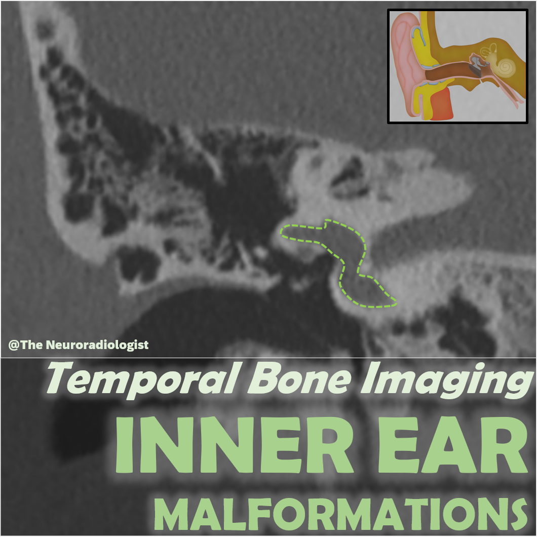
Temporal Bone Imaging (3): Inner Ear Malformations
On Sale
€3.99
Pay what you want:
(minimum €3.99)
€
This package contains the slides from the video "Imaging of Inner Ear Malformations" in three PDF formats:
- Full-size slides – One slide per page, great for viewing on screen
- Handout version – Six slides per page, ideal for quick review
- Note-taking version – Three slides per page with space for your own notes
A detailed case-based guide to imaging of inner ear malformations, including anatomy, embryology, and the full range of congenital abnormalities. Topics include complete labyrinthine aplasia, common cavity, cochlear aplasia, the spectrum of cochlear hypoplasia (types 1–4), incomplete partition (types 1–3), enlarged vestibular aqueduct, cochlear nerve deficiency, internal auditory canal stenosis, and semicircular canal malformations. Essential for radiologists and ENT clinicians involved in pediatric imaging and cochlear implant work-up.
👉 Watch the full video here: Temporal Bone Imaging (3): Inner Ear Malformations. - YouTube