

Temporal Bone Imaging (1): Outer Ear Disease
On Sale
€3.99
Pay what you want:
(minimum €3.99)
€
This package contains the slides from the video "Imaging of Outer Ear Disease" in three PDF formats:
- Full-size slides – One slide per page, great for viewing on screen
- Handout version – Six slides per page, ideal for quick review
- Note-taking version – Three slides per page with space for your own notes
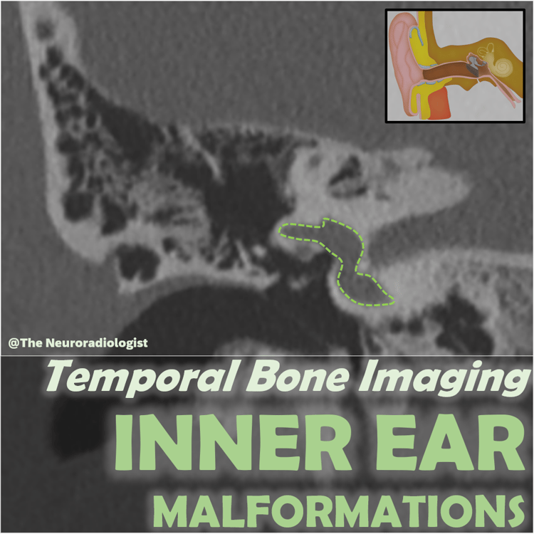
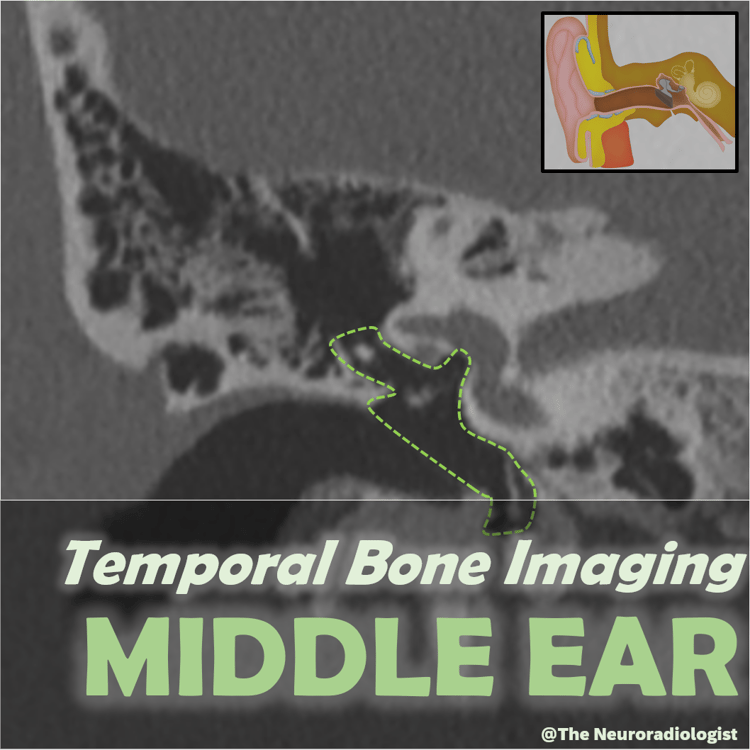
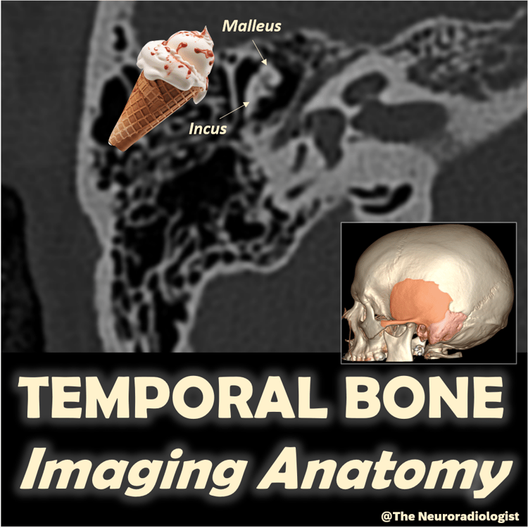
A detailed and case-rich overview of outer ear pathology on imaging. Topics include external auditory canal (EAC) atresia, necrotizing external otitis, EAC cholesteatoma, medial canal fibrosis, keratosis obturans, exostosis and osteoma, malignant neoplasms of the EAC, and eardrum pathology. This presentation combines anatomy, key pathology patterns, and essential diagnostic tips — highly relevant for radiologists and ENT specialists alike.
👉 Watch the full video here: Temporal bone Imaging (1): Outer Ear Disease