

Introduction to the Equine Obturator Nerve
Introduction to the Equine Obturator Nerve
Nobody talks about this one. Which is why so many horses get treated for hock problems they don't actually have.
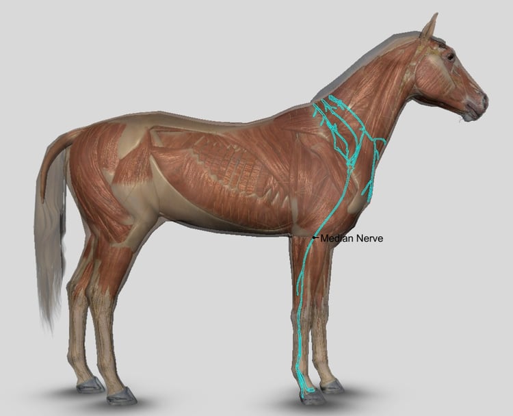
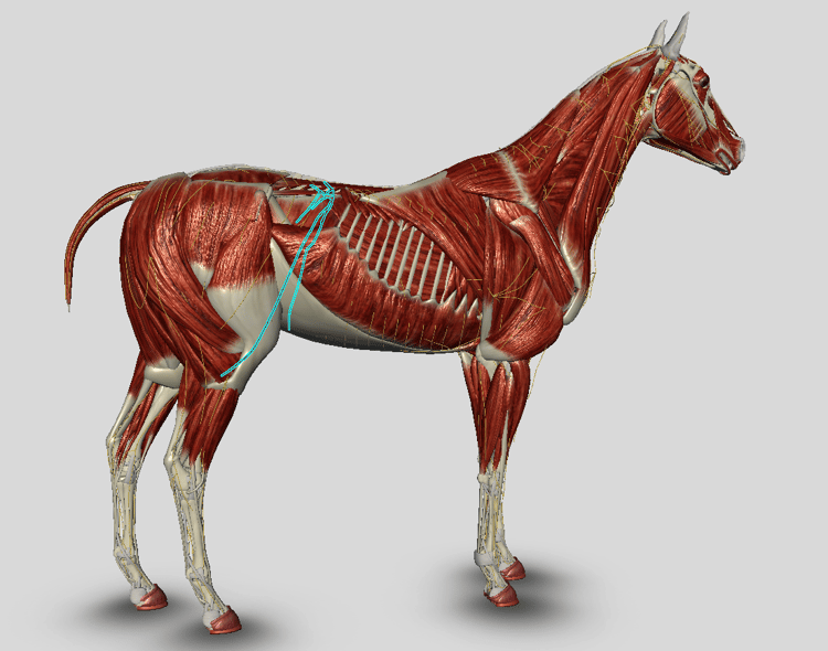
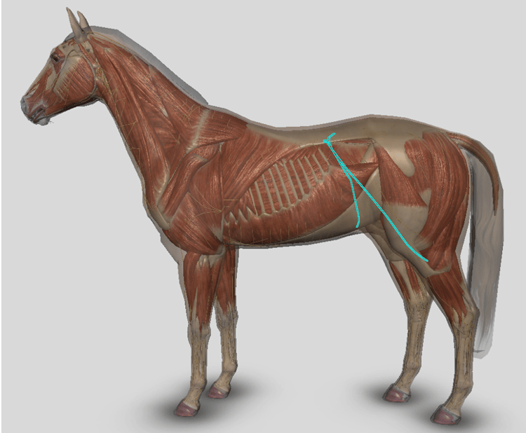
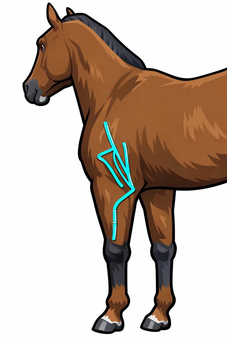
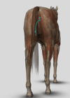
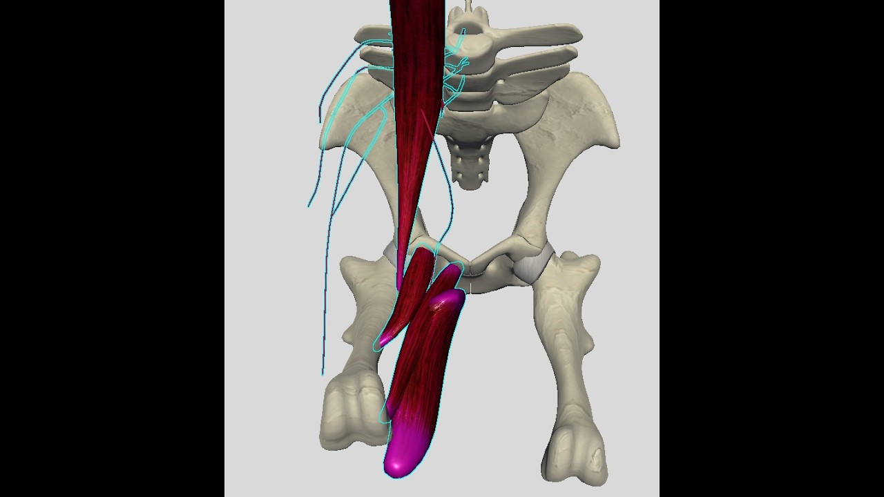
The obturator nerve exits the pelvis through the obturator foramen and supplies the adductor group — the muscles responsible for medial stabilization of the hindlimb during swing and early stance. When it's compromised, medial stabilization is delayed. The hock arrives slightly outside the ideal load line. It wobbles. And everyone looks at the hock.
The obturator nerve is reporting a lumbar problem through the medial hock. The hock is not the problem.
This nerve sits in the L4–L6 lumbar cascade, which means when the thoracolumbar hinge is compressed and the lumbar chain is loaded, the obturator is downstream. The adductors drop out. The limb loses its medial guide rail. The horse compensates — usually through the biceps femoris and caudal thigh — and that compensation gets labeled as stiffness, resistance, or a hock that needs injecting.
This webinar covers:
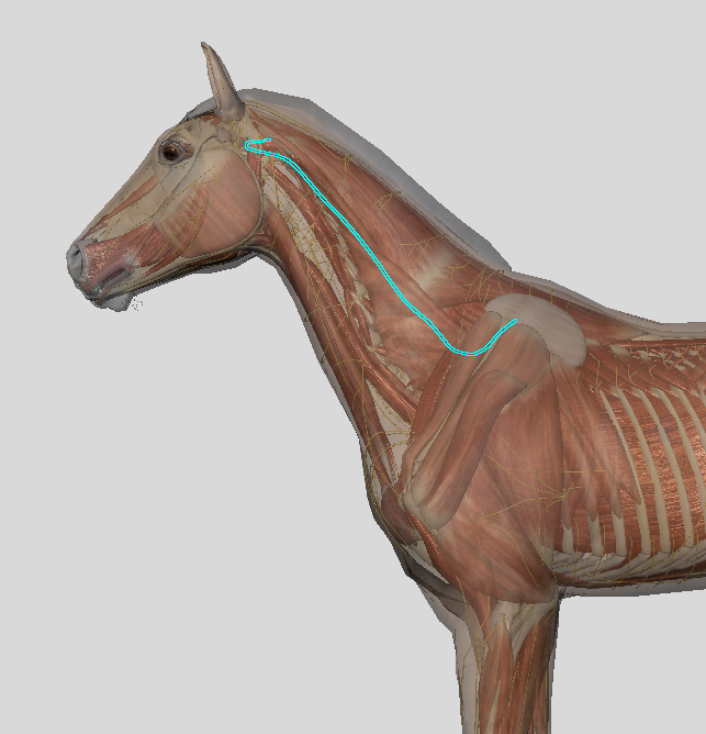
→ Where the obturator nerve is, where it goes, and what it controls
→ Why adductor dropout produces hock presentation
→ Its place in the lumbar cascade and what's driving it upstream
→ How to recognize the pattern clinically
→ MFR and related approaches for the obturator pathway
If you've got a horse with inside hind leg instability, hock wobble, or a thigh that won't release — start here.
Saturday July 11, 2026 10:00 AM Eastern USA
Replay is included with this webinar.